
FIGURE 5.
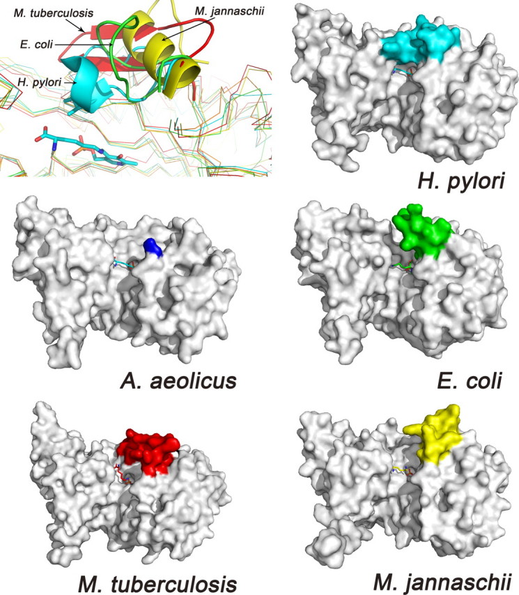
The active site loops of various DAPDCs. In the first panel, the five monomer structures are overlaid, and the active site loops are drawn as ribbon. In the next five panels, the structures are shown as surface, and the active site loops are colored differently. The ligand in AaDAPDC is taken from HpDAPDC to indicate the active site.