
FIGURE 5.
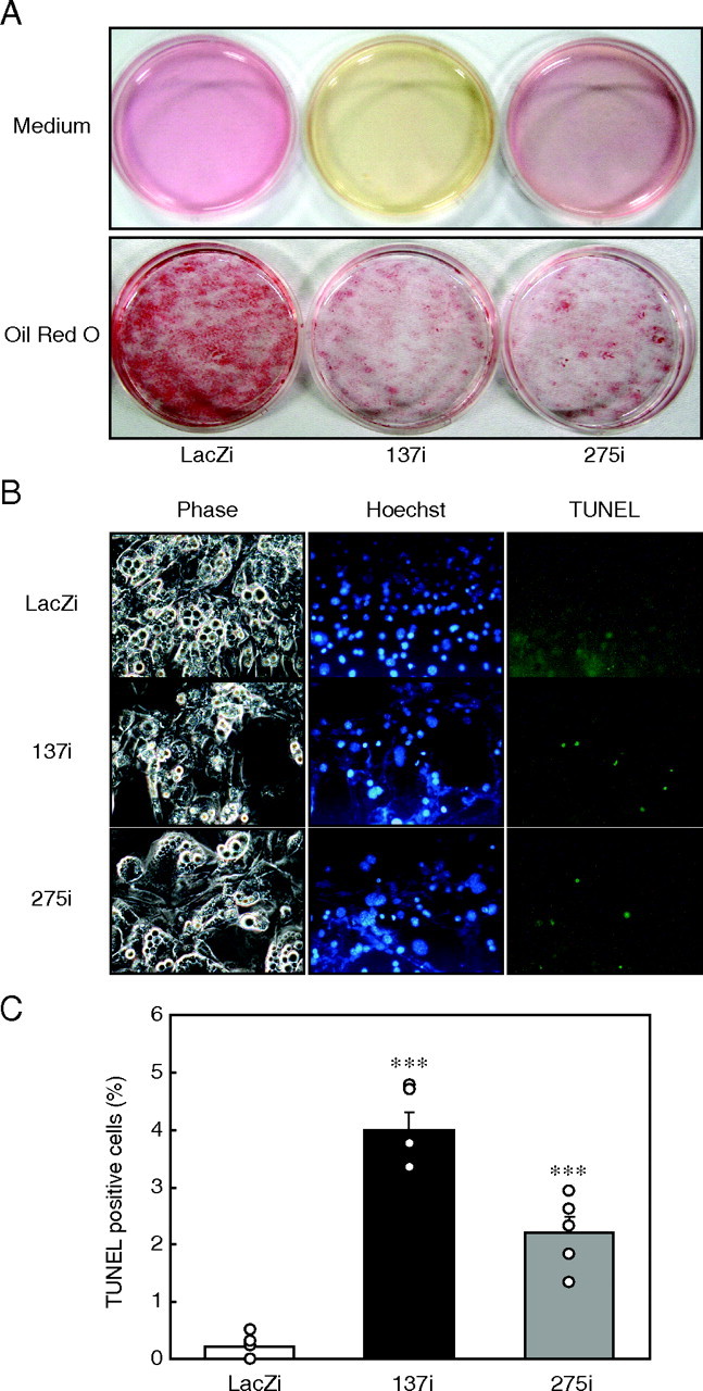
p21 gene knockdown induces apoptosis in 3T3-L1 adipocytes. A, 3T3-L1 cells were differentiated into adipocytes, and p21 RNAi adenovirus was infected at day 6. Media (upper panel) and Oil Red O staining of 3T3-L1 cells (lower panel) 4 days after infection of p21 RNAi (137i or 275i), or control LacZi adenovirus (day 10). B, apoptosis of 3T3-L1 adipocytes after p21 knockdown at day 10. Microscopic examinations of the cells with light phase contrast for morphology (phase (left)), Hoechst 33342 for nuclear staining (Hoechst (middle)), and TUNEL staining for apoptotic cells (TUNEL (right)) are shown at a magnification of ×400. C, emergence of apoptotic cells was determined by the ratio of TUNEL positive cells to Hoechst 33342-positive cells. Values represent the mean ± S.E. from six dishes per group. ***, at p < 0.0001 for 137i and 275i versus LacZi adenovirus-infected cells.