Figure 5.
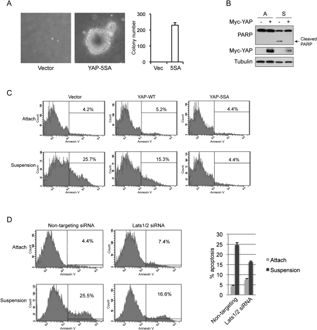
YAP inhibits detachment-induced anoikis. (A) Active YAP induces anchorage-independent growth in MCF10A cells. MCF10A cells infected with control vector or YAP-5SA were cultured in soft agar for 16 d before pictures were taken. Colony number was counted and is shown in the right panel. (B) YAP inhibits suspension-induced PARP cleavage. Control or YAP-expressing HMLE cells were cultured on tissue culture plates (A) or ultralow attachment plates in suspension (S) for 72 h. Cells were then harvested and analyzed by Western blotting with specific antibodies. (C) YAP inhibits anoikis. HMLE cells with control vector or ectopic expression of YAP wild type or 5SA mutant were cultured on tissue culture plates (Attach) or ultralow attachment plates in suspension for 72 h. Cells were then collected and stained with PE Annexin V and analyzed by FACS. Experiments were done in duplicate. (D) Knockdown of Lats1/2 inhibits anoikis in HMLE cells. HMLE cells were transfected with control siRNA or siRNAs targeting Lats1 and Lats2. Cells were then cultured on tissue culture plates (Attach) or ultralow attachment plates in suspension. After 72 h, cells were collected and stained with PE Annexin V and analyzed by FACS. Apoptosis rates were quantified from two independent experiments and are shown in the right panel.

