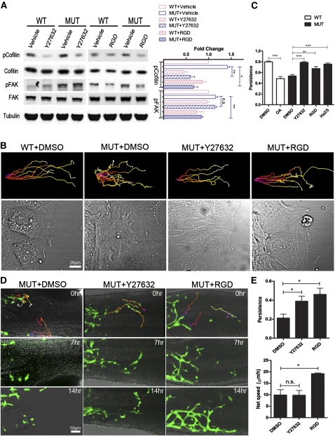
Figure 7.
Rescue of random migration both in vitro and in vivo. (A) MEFs were grown on laminin and then wounded for 1 h. Where indicated, 5 μM Y27632, 100 μg/mL GRGDTP, or vehicle control was included during the wound healing period. Cells were lysed, and the cellular content of pSer3-cofilin, phospho-Y925-FAK, and β-tubulin relative to total cofilin and FAK was determined by Western blotting. Quantification of protein expression based on experiments such as shown. n = 3; (*) P < 0.05; (**) P < 0.01. (B) Confluent MEF monolayers were wounded, and the cells were allowed to migrate into the wound in the presence of 5 μM Y27632, 100 μg/mL GRGDTP, 10 μg/mL β1 integrin blocking antibody (Ha2/5 clone, Supplemental Movie S17), or vehicle control (DMSO). The cells were imaged every 3 min for 15 h, and then tracked by Imaris software. Representative trajectories of migrating cells (top panels) and selected phase-contrast images showing lamellipodia morphology of migrating cells (bottom panels). (C) Quantification of cell persistence. n > 300 track plots; (**) P < 0.01; (***) P < 0.001. Data are expressed as mean ± SD. (D) Still images from time-lapse movies of E12.5 Phactr4humdy/humdy;RetTGM/+ hindgut explants treated with DMSO, 20 μM Y27632, or 1 mg/mL GRGDTP and then imaged every 3 min for 16 h. Time is noted in hours. (Top panels) Cell trajectories were color-coded to indicate the relative time point. (E) Quantification of cell persistence and net speed. (*) P < 0.05. Data are expressed as mean ± SD in three independent experiments.

