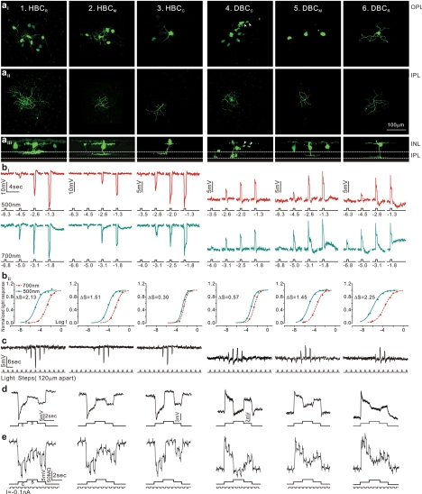
Figure 4.
Morphology, light responses, and receptive fields of six types of bipolar cells in the tiger salamander retina. (a) Fluorescent micrographs of a neurobiotin-filled HBCR (column 1), a HBCM (column 2), HBCC (column 3), a DBCC (column 4), a DBCM (column 5), and a DBCR (column 6) viewed with a confocal microscope at the outer INL/OPL level (ai), the IPL level (aii), and with z-axis rotation (aiii). Calibration bar, 100 μm. (bi) BC voltage responses to 500-nm and 700-nm light steps of various intensities. (bii) Response-intensity (V-Log I) curves of the responses to 500-nm and 700-nm lights. ΔS (spectral difference, see Fig. 3A) of the 6 BCs are 2.13, 1.51, 0.30, 0.57, 1.45, and 2.25. (c) Measurements of BC receptive field center diameters (RFCD) by recording voltage responses to a 100-μm-wide light bar moving stepwise (with 120-μm step increments) across the receptive field. (d) Voltage responses of the 6 types of BCs elicited by a center light spot (300 μm) and a surround light annulus (700 μm inner diameter, 2000 μm outer diameter). The surround light annulus was of the same intensity (700 nm, −2) for all 6 cells whereas the intensity of the center light spot was adjusted so that it allowed the annulus to produce the maximum response. (e) Voltage responses of the 6 types of BCs elicited by a center light spot and a surround light annulus (same as in d), and by a train of −0.1-nA/200-msec current pulses passed into the cell by the recording microelectrode through a bridge circuit. (Reprinted with permission from Zhang A-J, Wu SM. Receptive fields of retinal bipolar cells are mediated by heterogeneous synaptic circuitry. J Neurosci. 2009;29:789–797. © 2009 by The Society for Neuroscience.)

