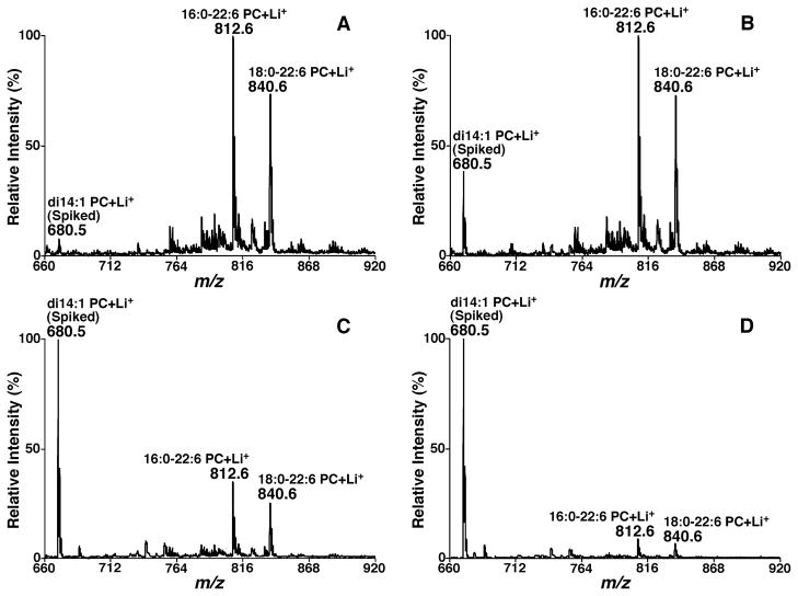
FIGURE 22.
Representative mass spectrometric analysis of a mouse myocardial lipid extract with different amounts of spiked di14:1 phosphatidylcholine. Different amounts of di14:1 phosphatidylcholine (PC) (0.16 pmol/μl in Panel A, 0.8 pmol/μL in Panel B, 4 pmol/μL in Panel C, and 16 pmol/μL in Panel D) prior to the MS analysis were spiked into a fixed amount of a lipid extract of mouse myocardium (approximately 6 pmol/μL), which was prepared in the absence of di14:1 PC during lipid extraction. Full mass spectra were directly acquired after infusion of this diluted solution in the presence of a small amount of LiOH (20 pmol/μL). The ion at m/z 680.4, which corresponds to lithiated di14:1 PC, is specified.

