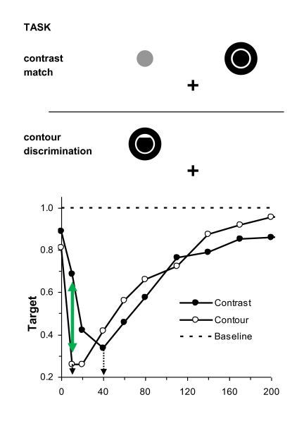
Figure 1.
Top panel: The spatial layout of stimuli used to study a target’s surface and contour processing during metacontrast. In the contrast matching task, the target and comparison stimulus were presented slightly above and symmetrically to the right and left of fixation, and only the target was followed by a mask. The observer on any trial had to indicate which of the two stimuli, target or comparison, appeared darker. In the contour discrimination task, the target and following mask were both centred slightly above and either to the left or else to the right of fixation. The target could be a disk with either an upper contour deletion (as shown), a similar lower contour deletion, or no deletion. Here the stimulus display position and target shape was randomized over trials. Using a three-alternative forced-choice procedure, on any trial the observer had to indicate which of the targets was presented. Bottom panel: The normalized target visibility functions, relative to a baseline visibility of 1.0 obtained when the targets were presented without the following mask, shown separately for surface contrast and for contours, as a function of the stimulus onset asynchrony (SOA) between the target and the mask. Note (a) the difference of 30 ms between the optimal masking obtained for the contour discrimination and the contrast matching tasks (dotted arrows) and (b) the dissociation between contour and contrast visibilities at the SOSOA of 10 ms (dashed arrow). Adapted from “Meta- and Paracontrast Reveal Differences Between Contour and Brightness-Processing Mechanisms” by B. G. Breitmeyer, H. Kafaligönül, H. Öğmen, L . Mardon, S. Todd, and R. Ziegler, 2006, Vision Research, 46, pp. 2646, 2647.

