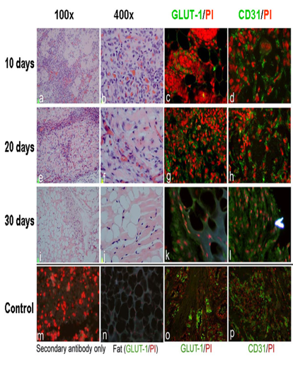
Figure 5.

In vivo hemangioma mouse model. Tissue sections of tumors formed after injection of cells from IH tumor spheres into mice were examined after 10, 20 and 30 days. (a, b, e, f, i, j) Hematoxylin and Eosin Staining (c, d, g, h, k, i) Immunofluorescent staining with anti-CD31 and anti-GLUT1 antibodies. Secondary antibody only (m) and mouse fat tissue staining with GLUT-1 (n) were used as negative control. Human hemangioma staining with GLUT-1(o) and CD31 (p) were used as positive controls.
