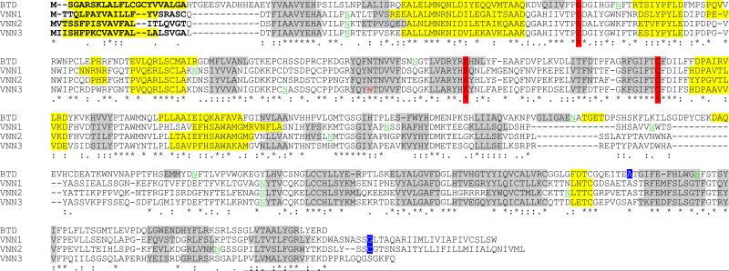
Figure 1.
Human Vanin family structural predictions. Predicted N glycosylation sites shown as underscored residues in green: N-Signal peptides in bold; predicted active site residues (based on biotinidase) boxed in red including * ; predicted α-helices in yellow (light shading); predicted β-sheets shaded in grey (dark); predicted GPI-anchor site residues in white with blue box; * identical residues; : conservative substitution; . non-conservative substitution; Missense residue in VNN3 shown in strikeout and red has been changed to allow readthrough to the predicted stop codon.

