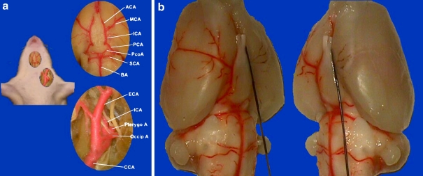
Figure 1.
Endovascular middle cerebral artery occlusion model in Sprague Dawley rats. a. Vascular anatomy of the left carotid artery system and Willis’ Circle. ACA: anterior cerebral artery. ICA: internal carotid artery. CCA: common carotid artery. ECA: external carotid artery. MCA: middle cerebral artery. PCA: posterior cerebral artery. PcoA: posterior communicating artery. SCA: superior cerebellum artery. BA: basilar artery. Occip A: occipital artery. Pterygo A: pterygopalatine artery. Note the potent posterior communicating artery in SD rat. b. With a 3–0 monofilament suture inserted from left ECA and gradually advanced into the intracranial ICA, blood flow was blocked to the left MCA territory

