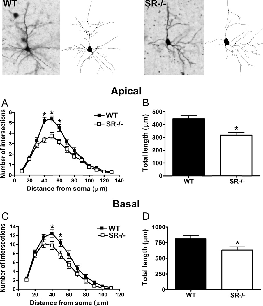
Fig. 1.
Pyramidal neurons in the somatosensory cortex of SR−/− mice display perturbations in apical and basilar dendritic morphology. The apical and basal dendrites of pyramidal neurons (3–8 neurons/subject) in the cortex from wild-type (WT; n= 6 mice; black) and SR−/− (n = 6 mice; white) animals were compared. Golgi stained pyramidal neurons and computer-assisted reconstructions of representative neurons in WT (left panel) and SR−/− (right panel) mice. (A), The apical dendrites of neurons from SR−/− mice were significantly less complex than WT mice between 40–60 µm from the soma. (B), The total dendritic length of neurons in SR−/− mice was significantly less than WT mice. The basal dendrites of neurons from SR−/− mice also showed significant reductions in (C), complexity (40–50 µm from the soma) and (D), total length. Asterisk (*) indicates significant difference from the WT group (p < 0.05). All values represent the mean ± SEM.

