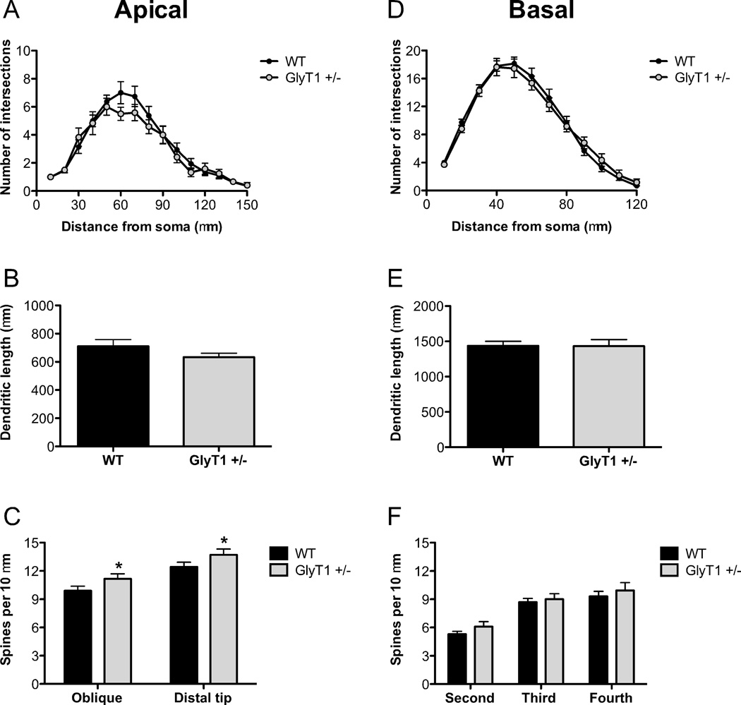
Fig. 7.
Pyramidal neurons in the somatosensory cortex of GlyT1+/− mice have increased apical dendritic spine density. The apical and basal dendrites of pyramidal neurons in S1 from wild-type (WT; n= 6 mice; black) and GlyT1+/− (n = 6 mice; gray) animals were compared. (A–B), There were no differences in the complexity of apical dendrites ((A)) or total apical dendritic length (B). (C), Spine density was increased in GlyT1+/− mice on both the distal portion and oblique branches of the apical dendrite. (D–F), There were no differences in the complexity of basal dendrites (D), total basal dendritic length (E), or spine density (F) of neurons from GlyT1+/− mice. Asterisk (*) indicates significant difference from the WT group (p < 0.05). All values represent the mean ± SEM.

