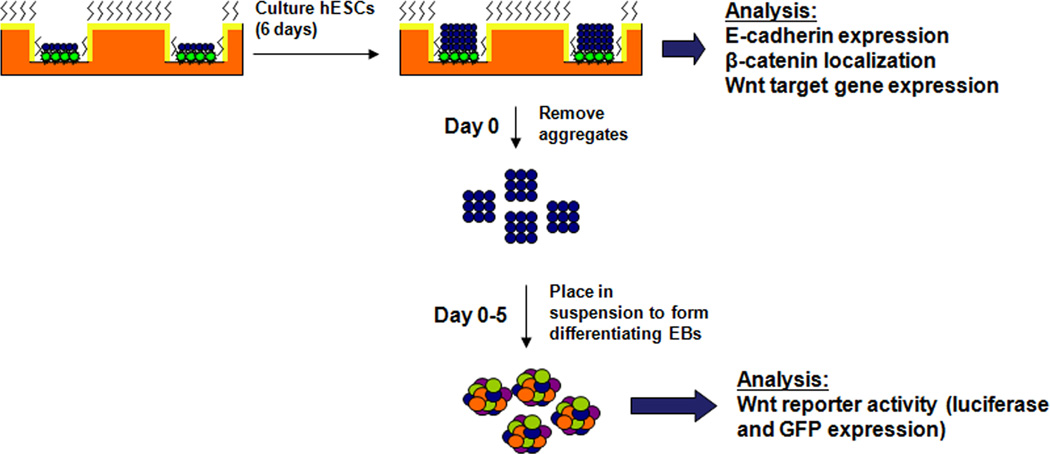
Figure 1. Schematic of microwell culture/EB differentiation process.
hESCs were cultured in 3-D microwells or on 2-D substrates for 6 days. At day 6 the cells were analyzed for E-cadherin expression, β-catenin localization, and Wnt target gene expression. For differentiation studies, on day 6 aggregates were enzymatically removed from either 3-D microwells or 2-D substrates and placed in suspension in medium containing serum. During the 5 day suspension period, Wnt reporter activity was assessed via assays for luciferase and GFP expression to quantify differences in Wnt signaling.

