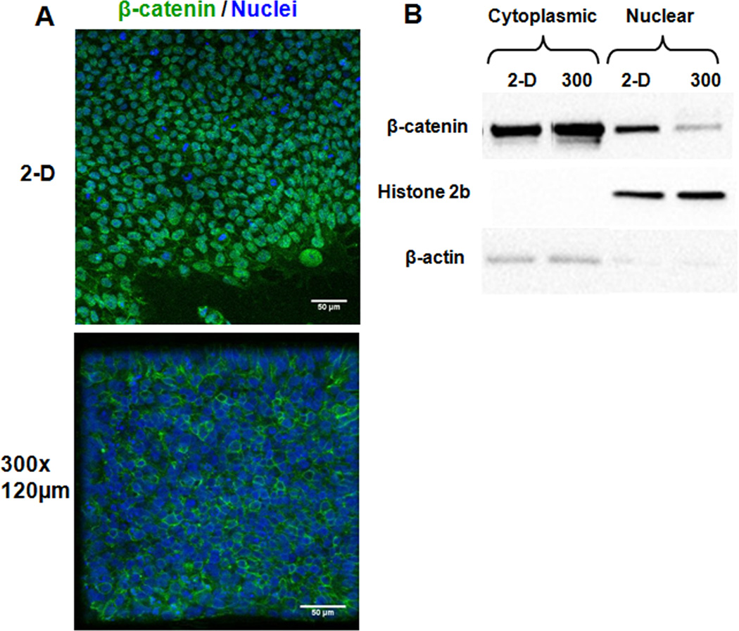
Figure 5. Microwell confinement of undifferentiated hESCs affects β-catenin localization.
Cells in 300×300×120 µm microwells and on 2-D substrates (Matrigel-coated glass coverslips) were labeled with an antibody for β-catenin (green) and a TOPRO-3-iodide nuclear stain (blue) at day 6 (A). Cells in microwells showed an absence of detectable nuclear β-catenin. This localization difference was confirmed via western blot analysis of nuclear and cytoplasmic protein extracts collected at day 6 from 300×300×120 µm microwells and 2-D Matrigel-coated TCPS plates (B).

