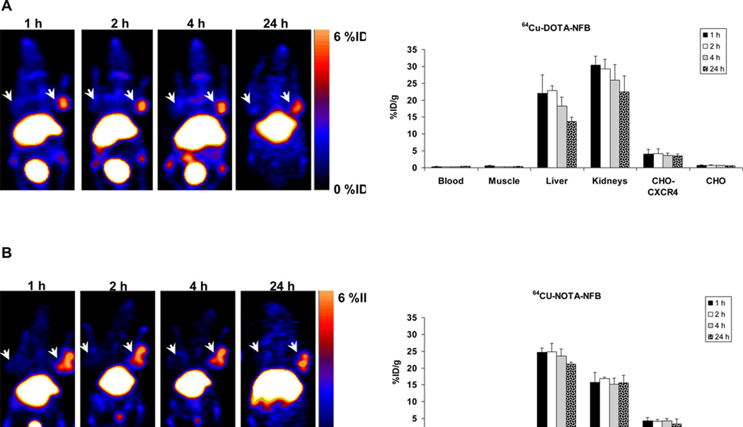
Figure 3.
(A) Representative coronal PET images (Left) and uptake calculation (Right) of mice injected with 100 µCi of 64Cu-DOTA-NFB (B) Representative coronal PET images (Left) and uptake calculation (Right) of mice injected with 100 µCi of 64Cu-NOTA-NFB. Arrows indicate CHO-CXCR4 tumor (right shoulder) and CHO tumor (left shoulder). Uptake results are calculated from PET scans and are shown as averages of 5–6 mice ± SE.

