
Figure 1.
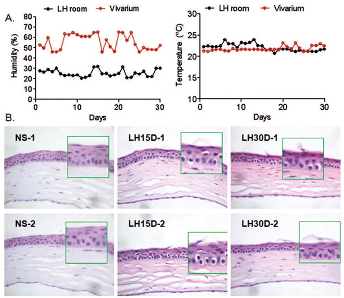
A. Mean relative humidity (%) and temperature (°C) rec ordings in the Low humidity room (LH) and vivarium throughout the experiment.
B. Representative H&E staining of corneas cryosections of C57BL/6 mice subjected to low humidity stress for 15 and 30 days (LH15D and 30D, respectively). Note desquamation of apical corneal epithelial cells at LH15D and 30D while control eye (nonstressed, NS) showed normal corneal architecture. Original magnification 10X. Insets indicate high magnification of left area immediately adjacent to inset.