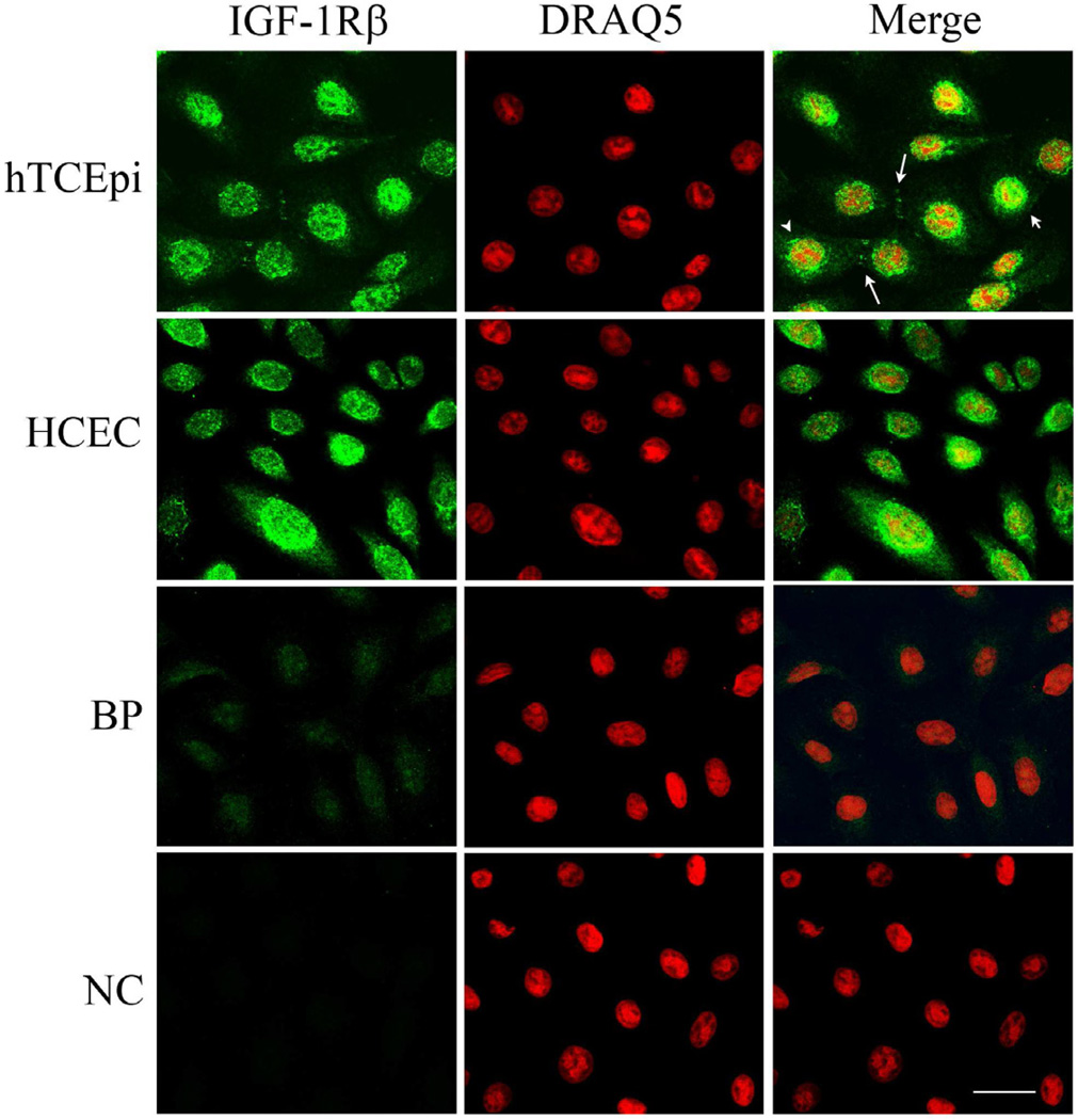
Figure 1.
Localization of IGF-1Rβ in corneal epithelial cells. IGF-1Rβ was detected in hTCEpi cells (first row) and HCEC cells (second row) using alexa fluor 488 (green, first column); nuclei were counterstained with DRAQ5 (red, second column); merged images (third column). Nuclear (small arrow) and juxtanuclear staining (arrow head) was noted in a variable distribution among cells. IGF-1Rβ was also detected at points of cell-cell contact (large arrow). A blocking peptide (BP, third row) was used to confirm antibody specificity. Negative control (NC, bottom row), primary antibody omitted. Scale: 22.5 µm.

