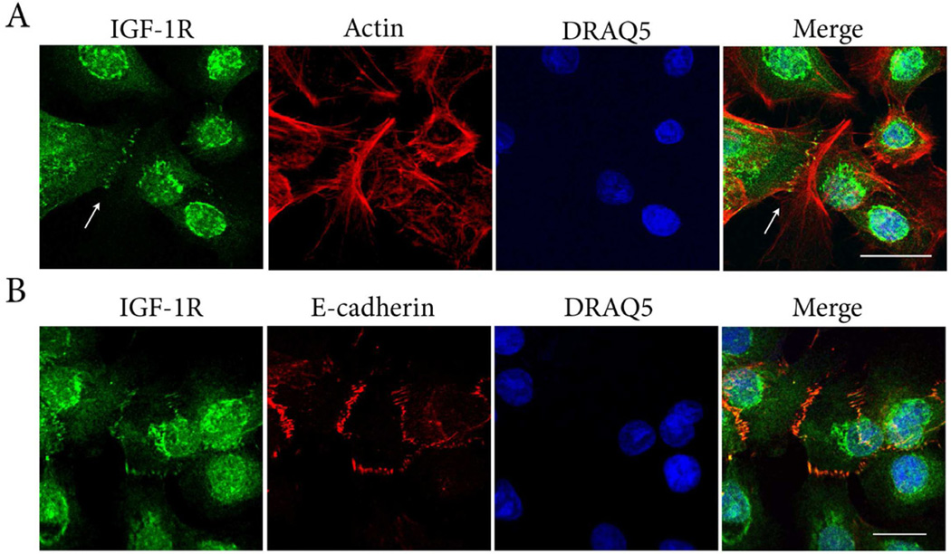
Figure 7.
IGF-1R:E-cadherin interactions. (A) Triple-labeling of hTCEpi cells with IGF-1Rβ (green), alexa fluor 543-phalloidin (red), and DRAQ5 stained nuclei (blue). Counterstaining with phalloidin demonstrated the presence of the IGF-1R localized to actin junctions at points of cell-cell contact (arrows). Scale: 28.73 µm. (B) Triple-labeling with IGF-1R (green), E-cadherin (red) and DRAQ5 stained nuclei (blue). IGF-1R at areas of cell-cell contact co-localized with E-cadherin (yellow). Scale: 17.12 µm.

