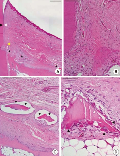
Figure 4.
Histological view of a poorly healed sample in the experimental group (H&E staining). (A) Low magnification. Arrowheads as in Fig. 2 (scale bar=1 mm). (B) Higher magnification at the notch area; minimal new bone was formed (scale bar=100 mm). (C) Fibrous encapsulated graft material (arrowheads; scale bar=250 µm). (D) Degradation of the residual graft material. Arrowheads, osteoclast-like cells (scale bar=50 µm).

