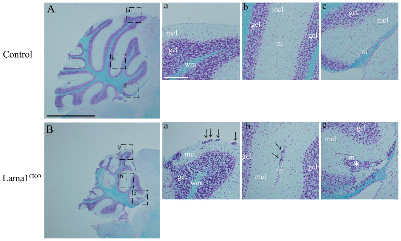
Fig. 2.
Cerebellar defects in Lama1CKO mice. Sagittal sections of adult control (A) and Lama1CKO mice at 8 weeks age (B) were stained with Luxol Fast Blue. The cerebellum of Lama1CKO mice is smaller than that of control mice, and the formation of some folia is disrupted. (a–c) High magnification view of areas indicated. In Lama1CKO mice, the aggregation of granule cells in the molecular layer was observed under meninges of the cerebellar surface and fusion lines of folia (arrows). The meninges, which form the fissure of the folia, were completely lacking (asterisk). mcl, molecular cell layer; gcl, granule cell layer; wm, white matter; m, meninges. Black scale bars: 1.0 mm, White scale bars: 100 μm.

