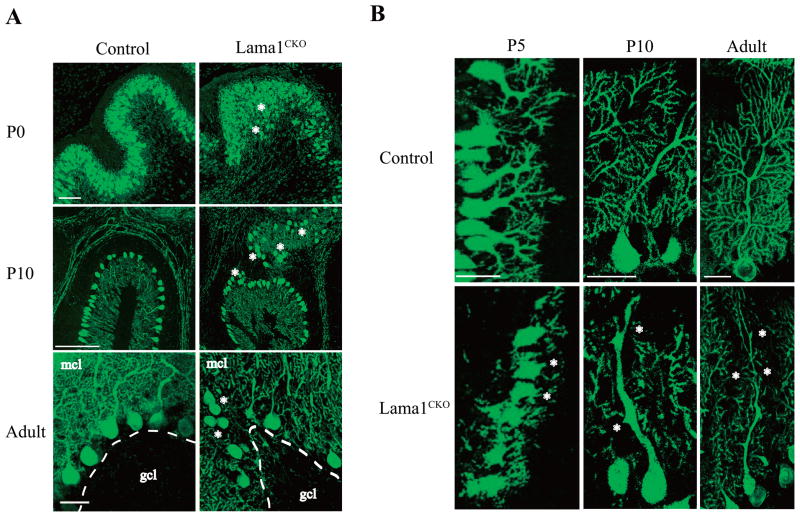
Fig. 3.
Abnormal arrangement and loss of processes in Purkinje cells in Lama1CKO mice. Brains from P0, P5, P10, and adult (8 weeks old) mice were cut into sections of 50 μm using a vibratome. The sections were stained with anti-calbindin D28K. Images were obtained every 2 μm at a thickness of 30 μm using a confocal laser microscope and staked using LSM510 software. (A) In the lobules of Lama1CKO mice, the arrangement of the Purkinje cell layer between the molecular layer (mcl) and granule cell layer (gcl) was disordered at P0, P10 and adult (asterisks). Scale bars: 50 μm. (B) The branching of dendritic processes of Purkinje cells decreased remarkably in Lama1CKO compared to control Lama1flox/del mice at P5, P10 and adult (asterisks). Scale bars: 50 μm.

