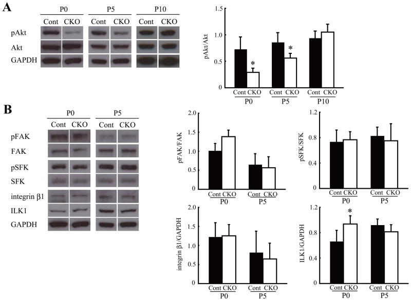
Fig. 5.
Reduced proliferation signaling in Lama1CKO mice. Cerebellum of control (Cont) and Lama1CKO mice (CKO) was analyzed by immunoblotting. (A) The phosphorylated Akt level was significantly decreased at P0 and P5 in Lama1CKO mice, compared with control mice, but this reduction was not observed at P10. (B) The expression of ILK1 was increased at P0 in Lama1CKO mice. The activation or expression of other integrin β related molecules was unchanged. The bar graphs show the mean and SD, which was calculated for each signaling density (n=5). (*, P < 0.05; two-sided t-test).

