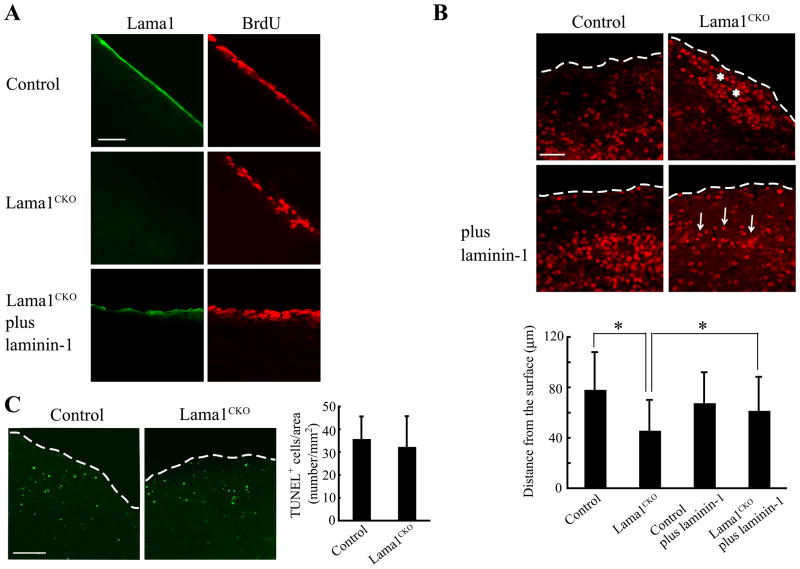
Fig. 6.
Reduced migration of proliferating granule cells in Lama1CKO mice. Sagittal slices of the cerebellum of P10 control and Lama1CKO mice were labeled with bromodeoxyuridine (BrdU) and then cultured. After culture, the slices were stained with anti-BrdU antibody and anti-Lama1. (A) At 1h after culture, lama1 was not expressed in the meninges in the slices of Lama1CKO mice, but the expression was recovered by adding 5 μg/ml laminin-1. Scale bars: 20 μm. (B) The migration of BrdU-positive granule cells to the inner layer in Lama1CKO mice decreased after a 72 h culture period (asterisks). However, the decrease could be rescued by adding 5 μg/ml laminin-1 (arrows). The dashed lines show the surface of the cerebellum. Scale bars: 20 μm. The bar graphs show the calculated mean and SD, (n=500 cells). (*, P < 0.001; two-sided t-test). (C) After culturing for 72h, the slices were stained using the TUNEL method. The dashed lines show the surface of the cerebellum. No differences were observed in the number of apoptotic cells in the control or Lama1CKO mice. The bar graphs show the calculated mean and SD. Scale bars: 100 μm.

