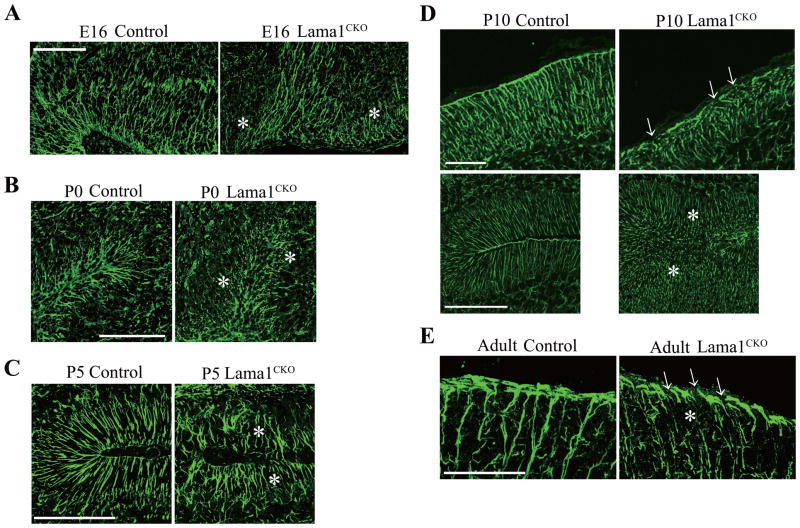
Fig. 7.
Aberration of radial and Bergmann glial fibers and endfeet in Lama1CKO mice. Cerebellar sections from E16 (A), P0 (B), P5 (C), P10 (D) and adult (E) mice were stained with anti-radial glial marker-2 or anti-GFAP antibody. In control mice, radial glia and Bergmann glial fibers extended to the meninges in the fissure of folia, whereas in Lama1CKO mice, the glial fibers were discontinuous and fragmented (asterisks). (D) At the surface of the cerebellum, Bergmann glial endfeet are continuous along the meninges in control mice. Some endfeet are disrupted in Lama1CKO mice (D and E, arrows). Scale bars: 100 μm. Scale bars: 200 μm (A and B), 50 μm (C and E), 100 μm (D).

