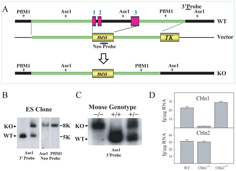
Fig. 6.
Generation of Cbln2-null mice. (A) Schematic representations of Cbln2 genomic DNA (WT), targeting vector (Vector) and targeted genome (KO). Shaded bars indicate the probes (neo and 3′) for Southern blot analysis. All 3 exons and introns of Cbln2 were replaced with a neo cassette. Southern blot analysis of genomic DNA was prepared from ES cells (B) and tissues of wild-type (+/+), heterozygote (+/−), and knockout (−/−) mice (C). DNA from ES cells and mouse tissues was digested with AseI (for 3′probe and neo probe) and PflMI (for neo probe), then analyzed using a DIG-labeled 0.25 kb 3′ external probe and a neo probe, respectively. Bands with the indicated sizes denote correct targeting in both ES cells and tissues. (D) qRT-PCR showing loss of Cbln1 and Cbln2 mRNA in Cbln1-null and Cbln2-null mouse brain, respectively. There is no compensatory change of Cbln1 or Cbln2 mRNA levels in Cbln2- or Cbln1-null mice, respectively.

