Fig. 3.
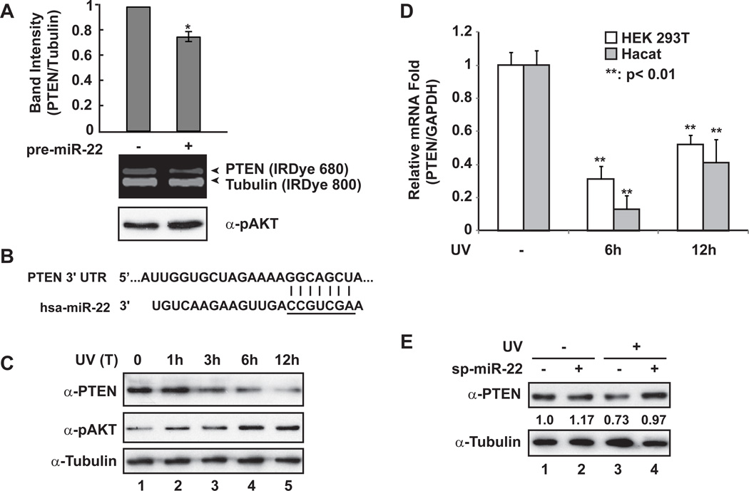
PTEN expression was repressed by miR-22 upon UV radiation. (A) HEK293 cells were transiently transfected with control or pre-miR-22. After 36 h, whole cell lysates were prepared and immunoblotted with anti-PTEN, anti-pT308 AKT and anti-Tubulin antibodies. Immunoblots were visualized with IRDye-labeled secondary antibodies as shown and quantified with Odyssey imagining system. *: p<0.05. (B) miR-22 recognition element within 3’-UTR of PTEN gene is complementary to miR-22 seed region (underlined). (C) HaCaT cells were treated with UV (20J/m2), and cells were harvested after treatment at the indicated times. Whole cell lysates were used for immunoblotting with antibodies against PTEN, pAKT and Tubulin. (D) HEK293T and HaCaT cells were treated with UV (20J/m2). PTEN and GAPDH mRNA level at 6 h or 12 h after UV treatment were analyzed with qRT-PCR, and the fold change of relative mRNA expression was shown as mean ± SD. (E) HEK293T cells were transfected with miR-22 sponge inhibitor as indicated. 36 h later, cells were mock treated or treated with UV (20J/m2). Cells were harvested at 6 h after treatment and whole cell lysates were subjected to immunoblotting with indicated antibodies. Immunoblot data were obtained and quantified as in (A).

