Fig. 3.
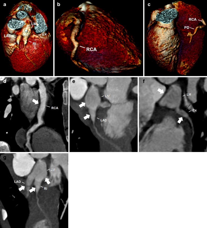
A 5-year-old boy with KD. Reconstructed volume-rendered 3D CT coronary angiography (a, b, c) revealing coronary arteries dilatations involving the right coronary artery (RCA), including the posterior descending artery (PD), the left main (LM) and the proximal segments of the left anterior descending artery (LAD) and circumflex artery (Cx) and the ramus intermedium (RI). Maximum-intensity projection 2D curved planar reformatting CT angiography of the RCA (d) showing diffuse ectasia (arrow) affecting all segments, including the PD. Maximum-intensity projection 2D curved planar reformatting CT angiography of the left coronary artery (e, f, g) revealing a giant coronary without intraluminal thrombus of the LM and proximal segments of the LAD, Cx and RI (arrows)

