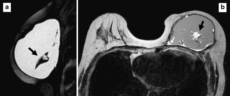
Fig. 11.
a Sagittal silicone-excited MRI sequence and (b) axial T2-weighted turbo spin-echo image of a 64-year-old woman with changes in the signal intensity of the silicone gel (black arrows). The margins of the implant are slightly irregular and a small amount of fluid surrounds the prosthesis (white arrow). A ruptured implant was confirmed at surgery.

