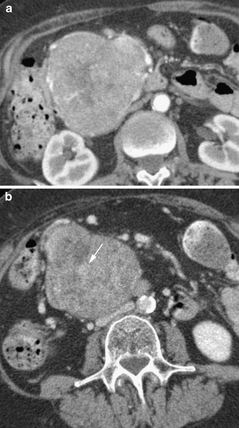
Fig. 5.
a Arterial phase CT in a 67-year-old woman with a heterogeneous soft tissue mass shown to be a microcystic cystadenoma. It almost appears solid with some central fluid attenuation elements. Note it displaces vessels at the periphery rather than invading and obstructing. b Portal venous CT in the same patient. Note eccentric central enhancing ‘scar’ (arrow)

