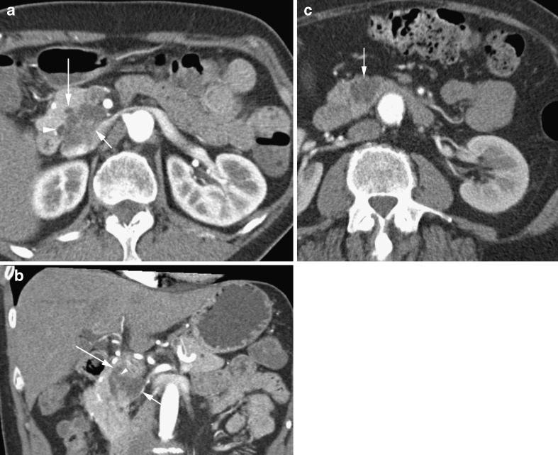
Fig. 7.
a Arterial phase CT in a 59-year-old man showing a lobulated cystic mass (short arrow) shown to be a side branch IPMN. Note pancreatic duct (long arrow). b Coronal reformat in the same patient showing the lobulated IPMN (short arrow) extending to the pancreatic duct (long arrow) via a short side branch (arrowhead). c Axial CT showing a lobulated cystic side branch IPMN in a 66-year-old woman

