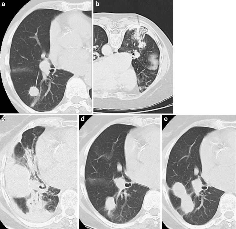
Fig. 4.
Primary lung tumour in the right upper lobe (A). The CT control during RFA demonstrates correct placement of the needle with a limited area of parenchymal haemorrhage (B). The CT control performed at the end of the procedure shows pleural effusion and a consolidation corresponding to parenchyma haemorrhage (C) without haemoptysis. No adjunctive procedures were required and the CT controls performed 3 (D) and 6 months (E) after treatment documented progressive reduction of the consolidation and shrinkage of the tumour nodule

