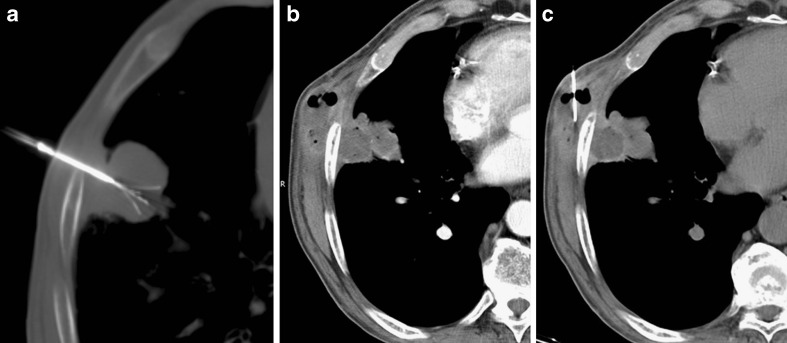
Fig. 5.
Subpleural lung tumour treated by RFA (A). The CT control performed 9 months after treatment depicted a subcutaneous mass adjacent to the tumour with peripheral enhancement and air bubbles. Under suspicion of a secondary location (tumour seeding), biopsy was performed (C), allowing the final diagnosis of an abscess formation involving the treated lesion with extension to the thoracic wall

