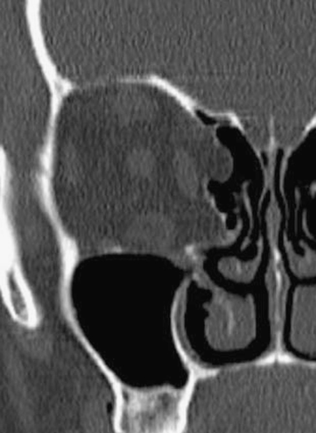
Fig. 10.

Two focal bone dehiscences are seen along the medial orbital wall, through which a small amount of orbital fat tissue herniates. This condition increases the risks of endoscopic sinus surgery, particularly if extrinsic ocular muscles are entrapped in the bone gap
