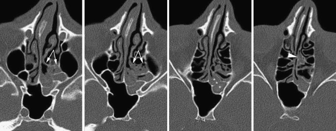
Fig. 12.
Sinusitis with a sphenoethmoid pattern: the sphenoethmoid recess is obstructed by thickened mucosa (asterisks), both the small left sphenoid sinus and the posterior ethmoid cells are opacified. Note the basal lamella (arrows) clearly demarcating the anterior ethmoid from the inflamed posterior ethmoid cells. Secretions are also retained within the pneumatised vertical lamella of the middle turbinate (arrowhead)

