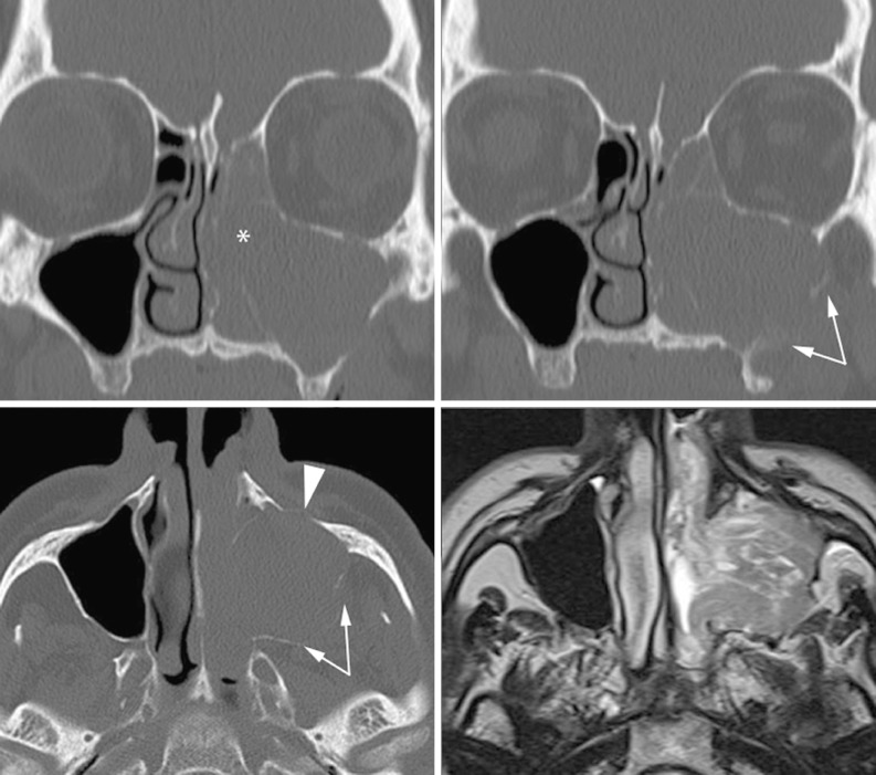
Fig. 20.
The left ostiomeatal complex is occupied by solid tissue, the anterior ethmoid cells, the maxillary and frontal sinus are completely opacified. The absence of inflammatory changes on the right side as well as the displacement and destruction of the anterior (arrowheadss) and the posterior (arrows) maxillary sinus wall suggests the presence of a neoplasm. Actually, MRI shows a mass arising from the maxillary sinus and protruding into the nasal fossa, histologically proven as ameloblastoma

