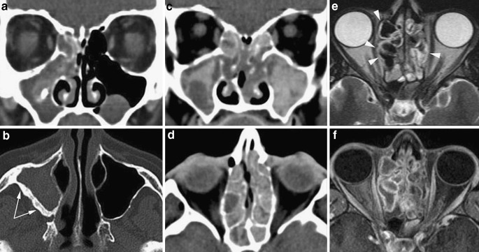
Fig. 21.
Non-invasive fungal rhinosinusitis. a, b The maxillary sinus is filled, at the periphery, by hypodense thickened mucosa and contains, in the centre, a calcification; dense sclerosis of the posterolateral wall secondary to chronic inflammation (arrows). These findings are consistent with a fungus ball. c, d Chronic rhinosinusitis with polypoid thickening of the mucosa and hyperdense material scattered in all sinus cavities: this pattern suggests allergic fungal rhinosinusitis. e, f On MRI eosinophilic mucin displays T2 hypointense signal comparable with air: the T1 intermediate signal along with bone remodelling and sinus expansion (arrowheads) help to make the diagnosis of allergic fungal rhinosinusitis

