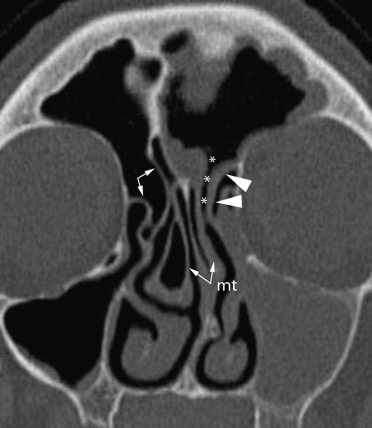
Fig. 3.
Coronal oblique MPR reconstruction showing the variable cranial attachment of the uncinate process. On the left side, the uncinate process attaches laterally to the medial orbital wall (arrowheads), thus the frontal recess (asterisks) courses close to the middle turbinate (mt). The ethmoid infundibulum is obstructed resulting in sinusitis with an infundibular pattern. On the right side, the uncinate process inserts on both the medial orbital wall and the skull base (arrows); the frontal recess (not visible) will drain into the middle meatus

