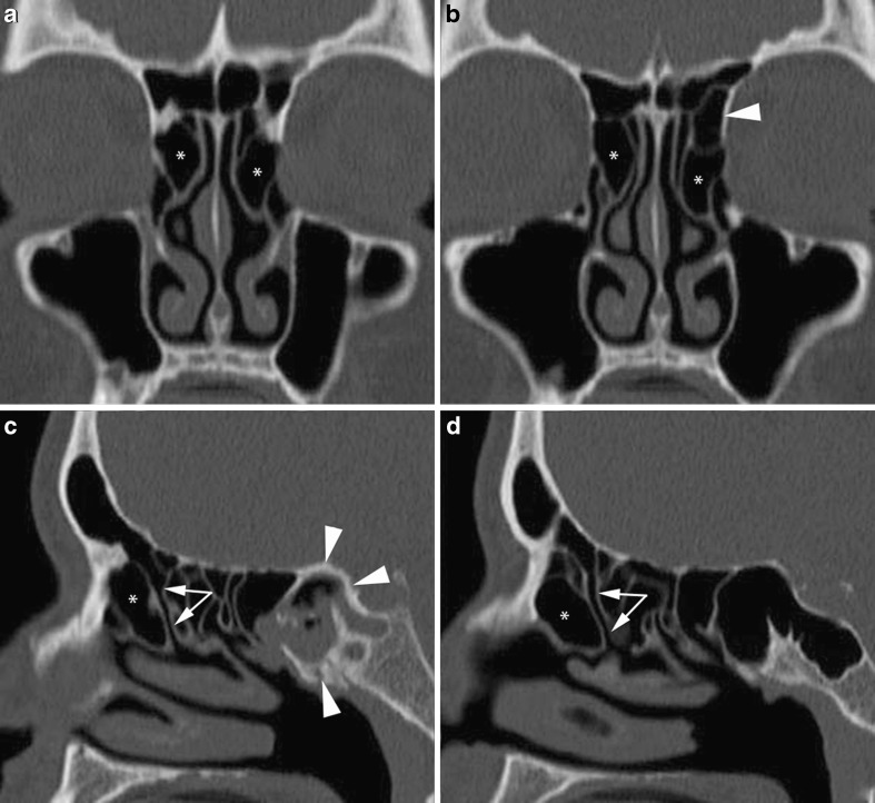
Fig. 5.
MSCT coronal (a, b) and sagittal reformations obtained on the right (c) and left (d) sides. Bilateral agger nasi cells (asterisks) are visible on the coronal images, but sagittal reformations better display their relationships with the frontal recesses (arrows). A type I bulla frontalis (arrowhead in b) can be seen on top of the left agger nasi cell. Inflammatory material with focal calcifications is retained within the right sphenoid sinus whose walls are densely sclerotic (arrowheads in c): chronic inflammation, possibly a fungus ball (see below)

