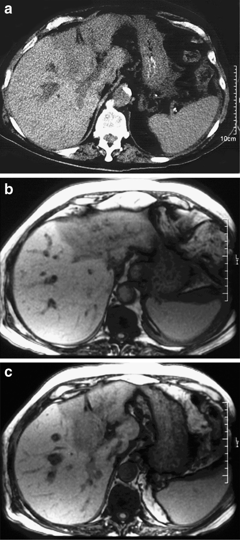
Fig. 5.
a–c A patient with left lobe atrophy associated with malignant hilar biliary obstruction due to metastasis of colorectal origin. The left portal vein branch is occluded. a Non-contrast-enhanced CT. The atrophied lobe shows lower attenuation than the non-atrophied lobe. b, c T1-weighted MRI. The atrophied left lobe shows lower signal intensity than the hypertrophied right lobe

