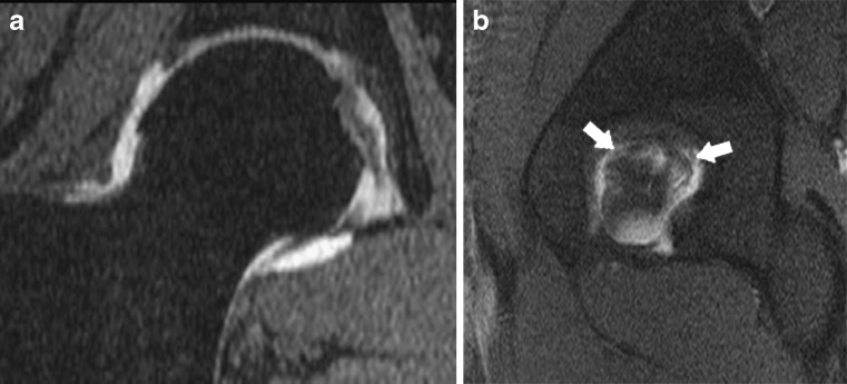
Fig. 15.
a, b Complete tear of the ligamentum teres in an 29-year-old man. a Coronal reconstruction from a fat-saturated 3D gradient echo acquisition: the acetabular insertion of the ligamentum teres is interrupted. The ligamentum teres has an abnormal intermediate signal. b Sagittal fat-saturated T1-weighted arthro-MR image through the acetabular bottom shows a laminated ligamentum teres completely vertically divided into two parts (white arrows)

