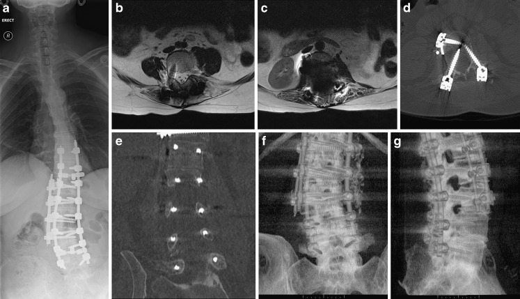
Fig. 11.
a–g Metal implants in the spine are frequently made of titanium. This does usually allow for satisfactory MRI. However, if ferrous metals or multiple implants are used MRI can become non-diagnostic. CT is usually still able to provide diagnostic images. In this example, two posterior and one lateral rod with corresponding anchoring screws have been used (a). Above the level of the lateral rod MRI results in diagnostic images (b), at the level of the lateral rod the artefact becomes too marked (c). CT imaging allows for excellent visualisation of the spine and implants and demonstrates misplacement of several pedicle screws (d, e). The coronal reformat in particular allows for a quick and accurate assessment of screw misplacement. Volume rendering allows for easy visualisation of implant placement in relation to the spine (f, g)

