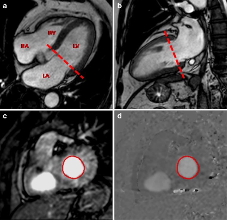
Fig. 1.
PC-CMR of transmitral inflow for assessment of diastolic function. Cine four-chamber (a) and LV vertical axis (b) showing the orientation of through-plane PC-CMR imaging (dashed line). (c) Magnitude and (d) phase-contrast images are obtained in a plane parallel to the mitral annular plane at the level of the mitral valve leaflet tips. The contour of the region of interest is drawn at the level of the leaflet tips and includes the whole cross-section of the transmitral inflow (red circle). LV, left ventricle; LA, left atrium; RV, right ventricle; RA, right atrium

