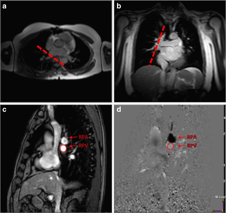
Fig. 2.
PC-CMR of pulmonary venous flow for assessment of diastolic function. Axial (a) and coronal (b) survey demonstrating the orientation of through-plane PC-CMR imaging (dashed line). (c) Magnitude and (d) phase-contrast images are obtained in a plane perpendicular to the right superior pulmonary vein (arrow), 1 cm away from the junction of the pulmonary vein with the left atrium. RPA, right pulmonary artery; RPV, right superior pulmonary venous

