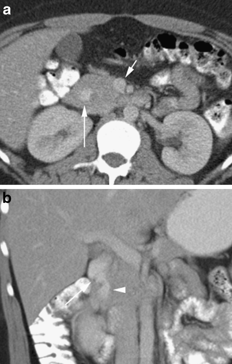
Fig. 13.
a Axial portal venous CT in a 22-year-old with a structure in the pancreatic head (long arrow) of similar density to the superior mesenteric vein (short arrow). b A coronal reformat shows this to be tubular (arrowhead) representing oral contrast agent within the second part of the duodenum in a patient with annular pancreas

