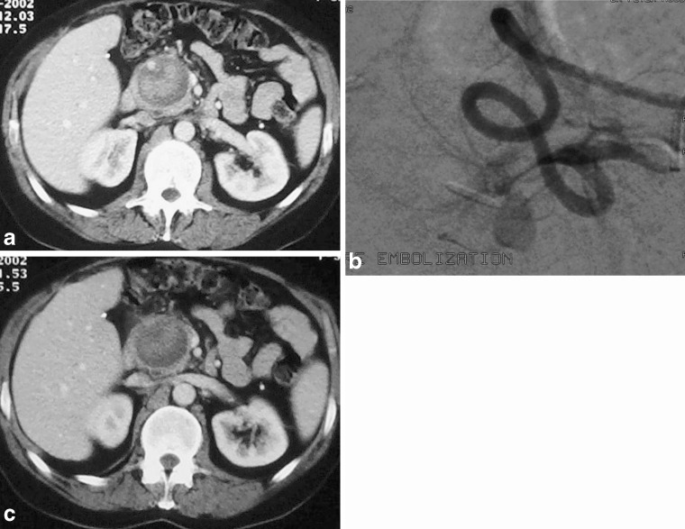
Fig. 8.
a Arterial phase CT showing a heterogeneous mass in the pancreas. Note the arterial enhancing lumen of the gastroduodenal artery at the 11 o’clock position in the pseudo-aneurysm in this patient with previous pancreatitis. b A DSA in the same patient clearly defines the pseudoaneurysm. c A portal venous phase at the same level. Note that the feeding artery is less well appreciated and the lesion could be mistaken for a pancreatic mass or collection

