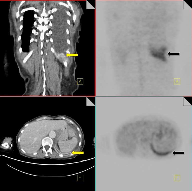
Fig. 12.
18F-FDG PET-CT performed in a 69-year-old male with a history of non-Hodgkin's lymphoma. The patient had a previous talc pleurodesis for a persistent left pleural effusion. Increased FDG activity is identified within the left pleura (black arrow). CT demonstrates a pleural effusion with high density material along the left pleural surface consistent with talc (yellow arrow)

