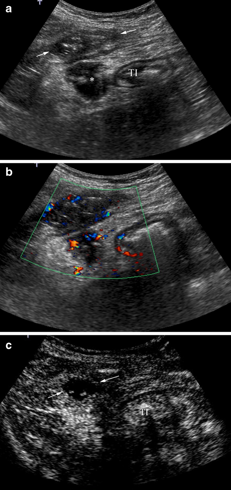
Fig. 12.
Anterior abdominal wall abscess. a B-mode US shows a thickened terminal ileum (TI) and two possible collections, in the anterior abdominal wall (arrows) and intraperitoneal (*). b Hypoechoic collections depict peripheral flow on colour Doppler. c Post-contrast agent image shows high enhancement of the intraperitoneal collection corresponding to a phlegmon (*). The collection located in the anterior abdominal wall is an area completely devoid of microbubble signal, representing an avascular abscess (arrows). CEUS defines its size better

