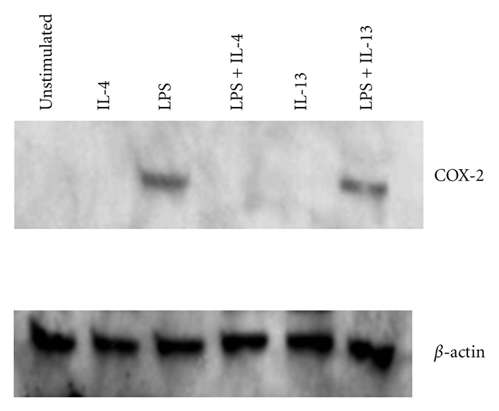
Figure 2.

COX-2 protein expression in resting and LPS-stimulated monocytes. Monocytes were isolated from blood by magnetic bead purification. Cells were treated with IL-4 (10 ng/mL), IL-13 (10 ng/mL), or LPS (1 μg/mL) for 24 hrs and whole cell lysates collected. Proteins were separated on a 10% SDS acrylamide gel and transferred to nitrocellulose. The membrane was probed with anti-COX-2 and then stripped and reprobed with anti-β-actin.
