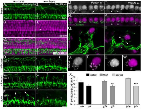
Figure 3. Defects in fasciculation in Pou3f4y/− mice impair innervation and synapse formation.
(A–L) Type I and Type II projections in Pou3f4y/+ and Pou3f4y/− embryos at E17.5. Only the distal 10 μm of the type I fibers are shown. Scale bar in L = 50 μm
(A–C) At the base of the cochlea, Type I fibers innervate the inner hair cell layer, while Type II fibers cross the pillar cell layer (shown in A and C) and then turn towards the base.
(D–F) Illustration of the base-to-apex gradient of maturation of type II fibers. At E17.5, Type II projections are abundant in the mid-base, sparse in the mid-apex, and short at the apex (arrows).
(G–I) In Pou3f4y/− embryos at E17.5, the Type I layer is less dense (see bracketed region), and there are fewer Type II projections (arrowhead).
(J–L) Compared with wild-type, Pou3f4y/− embryos show fewer, less mature Type II projections. At the apex (L), no Type II projections are observed. Scale bar = 40 μm.
(M–P) Whole-mount preparation of the apex of Pou3f4y/+ and Pou3f4y/− cochleae at P8. Anti-Ribeye immunostaining indicates ribbon synapses (see arrow in M). This antibody also recognizes CtBp2 (in all hair cell nuclei; n). IHC: inner hair cell. Anti-GluR2/3 (as in N) indicates post-synaptic glutamate receptors. Immunostaining for both factors in the mutant appears reduced (arrowheads).
(Q and R) Mid-modiolar cross-sections of the inner hair cell region from Pou3f4 y/+ (Q) and Pou3f4y/− (R) cochleae at E17.5. Nerve terminals are marked by anti-neurofilament (200 kDa). Note the decreased density of nerve fibers contacting the inner hair cell in Pou3f4y/−(arrowheads).
(S and T) A high-magnification view of the synaptic region from the inner hair cell in Q. Punctate spots (circled in T) represent individual ribbon synapses.
(U and W) Similar view as in S,T but from the Pou3f4y/− inner hair cell in R. Note the decreased number of ribbon puncta. Scale bar = 4 μm.
(X) Histogram indicating a significant decrease in the number of inner hair cell synapses (mean +/− SEM) in Pou3f4y/− cochleae at P8. **P ≤ 0.01; ***P ≤ 0.001.

