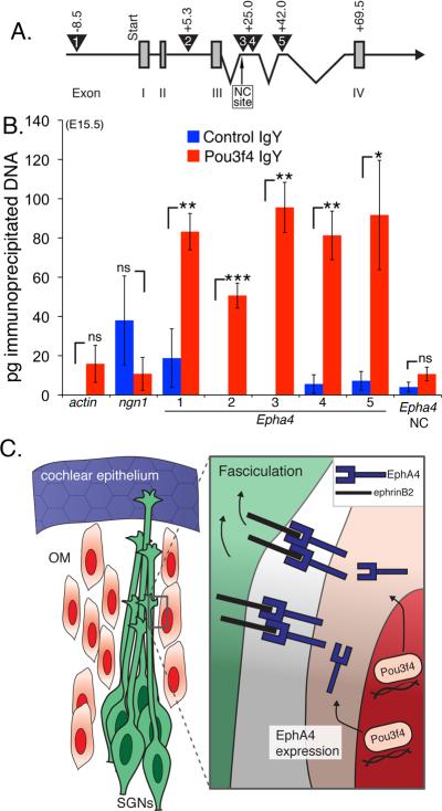
Figure 8. Pou3f4 protein associates with Epha4 regulatory elements.
(A) A cartoon schematic of Epha4 showing the first four of 16 exons. Exons I–IV (numbered gray boxes). The numbered triangles indicate the regions containing the putative Pou3f4 binding site ATTATTA. Primer sets were designed to amplify these five regions after ChIP. The negative control site, “NC site.”
(B) Histogram showing the number of picograms (pg) of immunoprecipitated chomatin for each primer set. ns = not significant. *P ≤ 0.05; **P ≤ 0.01; ***P ≤ 0.001. Mean +/- SEM.
(C) Cartoon schematic of a proposed model. Pou3f4 in otic mesenchyme induces expression of Epha4/EphA4, which binds to ephrin-B2 on developing SGN axons leading to fasciculation. As a result, SGN axons fasciculate to give rise to the inner radial bundles.

